Rv Lv Ratio Pe Radiology

Amgrad Rv Lv Ratio

Measurement Of Rv Lv Ratio Axial Ct Images Demonstrate The Best Download Scientific Diagram
Http Pdf Posterng Netkey At Download Index Php Module Get Pdf By Id Poster Id 127655

Right Heart Strain Radiology Reference Article Radiopaedia Org

Rv Lv Diameter Ratio 1 Not Associated With Worse Outcomes In Acute Pe Pulmonology Advisor

Gorgeous Rvot Ultrasound Humor Ultrasound Technician Medical Photos
Measurement made on axial imaging.
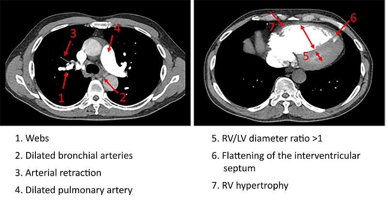
Rv lv ratio pe radiology. However incremental value of rvef over axial rv lv ratio was not found. Ratio 0 9 considered positive. Additional studies have estimated that an rv lv diameter ratio superior to 1 5 indicates a severe episode of pe 36 39 41. Ischemic congenital valvular heart.
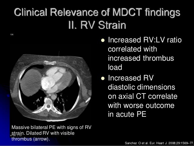
Rvef and rv lv ratio proved better predictors for outcome than pulmonary obstruction index both p 001. To retrospectively determine whether three computed tomographic ct findings ventricular septal bowing vsb ratio between the diameters of right ventricle rv and left ventricle lv and embolic burden are associated with short term death defined as in hospital death or death within 30 days of ct whichever was longer due to acute pulmonary embolism pe. The study authors found that greater clot volume was associated with a higher incidence of right heart dysfunction and that a right ventricle rv to left ventricle lv ratio that is greater. In the study by araoz et al 42 an rv lv diameter ratio greater than 1 was associated with a 3.
Rvef was the best predictor for clinical outcome in patients with acute pe. This retrospective cohort study included 579 consecutive subjects 08 2003 03 2010 diagnosed with acute pe with normal ct rv lv ratio 0 9 236. Although the prevalence of rv dysfunction defined as a right to left ventricular rv lv diameter ratio 0 9 was lower than in previous studies for example 66 in unselected and 63 in normotensive pe patients a prevalence of 54 appears considerably high in an apparently healthy and haemodynamically stable cohort of pe patients. The objective of this study is to identify a clinical scenario for which normal ct derived right to left ventricular rv lv ratio is sufficient to exclude rv strain or pe related short term death.
Patients with interstitial lung disease ild may develop pulmonary hypertension ph often disproportionate to the severity of the ild. Measurements will likely be on different axial images to obtain the true maximum measurement for each ventricle. The right ventricular to left ventricular diameter rv lv ratio measured at ct pulmonary angiogram ctpa has been shown to provide valuable information in patients with pulmonary arterial hypertension and to predict death or deterioration in acute. Right ventricular dysfunction usually results from either pressure overload volume overload or a combination.

Echocardiography In Pulmonary Embolism The Clot Thickens Pulmonary Embolism Pulmonary Pulmonary Emboli

Pin By Earl On Work Cardiac Sonography Diagnostic Medical Sonography Echocardiogram
Plos One Accuracy And Reproducibility Of Ct Right To Left Ventricular Diameter Measurement In Patients With Acute Pulmonary Embolism

Apical Hypertrophic Cardiomyopathy A Lv Angiography Demonstrates Apical Hypertrophy Hypertrophic Cardiomyopathy Human Body Anatomy Diastolic Heart Failure
Https Journal Chestnet Org Article S0012 3692 19 31374 1 Pdf

Apical 4 Chamber View Tee Diagnostic Medical Sonography Medical Ultrasound Cardiac Sonography

Tee Echocarigraphy Echocardiography Central Lakes Medical In 2020 Medical Photos Medical Central Lake

Https Encrypted Tbn0 Gstatic Com Images Q Tbn 3aand9gctdtuy2pnbihwpvwrua8qmba3h5lcihlvpnga Usqp Cau

I Impaired Relaxation Ii Moderate Diastolic Dysfunction Pseudonormal Iii Restrictive Left Ventricular Filli Cardiac Sonography Echocardiogram Cardiology

Imaging Right Left Ventricular Interactions Jacc Cardiovascular Imaging

Pin On Lub Dub 3

Pin By Brooke Kelso On Echo Pulmonary Embolism Diagnostic Medical Sonography Cardiac Sonography

8 Tips To Correct Rv Function Assessment With Tapse S Wave S Wave Cardiac Sonography Assessment

Ultrasound Registry Review Valvular Abnormalities Valvular Cardiac Sonography Ultrasound Sonography

73bc5cdb5b9e1af9a875461c4ae4a391 College Life Ultrasound Jpg 720 554 Medical Ultrasound Echocardiogram Cardiac Sonography
Imaging Of Pulmonary Embolism

Pin By Bauyrzhan Erbolatov On Geriatrics Internal Medicine Medical Photos Cardiac Sonography Echocardiogram

Use Of The Echocardiogram To Define The Presence Extent And Etiology Of Cardiac Dysfunction Echocardiogram Cardiac Sonography Cardiac
Https Encrypted Tbn0 Gstatic Com Images Q Tbn 3aand9gcsmnit Cbktx7mceehchohiwmhirjkzwqfopwqqwwqiml8g5yti Usqp Cau

Qp Qs Ratio In Echo Echocardiography Barnard Health Care In 2020 Echocardiogram Ductus Arteriosus Ventricular Septal Defect

Pin By Andres Sanchez On Cardiology Echocardiogram Diagnostic Medical Sonography Cardiac Sonography

Measurement Of Right And Left Ventricular Diameter Download Scientific Diagram

Pin On Structure And Function
Http Pdf Posterng Netkey At Download Index Php Module Get Pdf By Id Poster Id 120662
External Validation Of Radiological Signs Of Chronic Thromboembolic Isth Academy Boon D Jul 9 2019 273891

Right Ventricle Dilation After Pulmonary Embolism Relevant Rv Dilation Download Scientific Diagram

Mitral Stenosis Cardiac Sonography Vascular Ultrasound Cardiology

Left Ventricle Segmentation Echocardiography In Icu Medical Ultrasound Cardiology Nursing Ultrasound

Pin On Education

Qp Qs Ratio In Echo Echocardiography Barnard Health Care Coronary Artery Disease Aortic Dissection Coarctation Of The Aorta

Pericarditis With Fluid Heart Ultrasound Medicin

The Flail Leaflet Mitral Valve Prolapse Mitral Valve Prolapse Mitral Valve Echocardiogram

Pin By Didi Ramos On Ultrasound Medical Ultrasound Echocardiogram Cardiac Sonography

Sign Up Cardiac Sonography Diagnostic Medical Sonography Echocardiogram

Pin On Echocardiography

Qp Qs Ratio In Echo Echocardiography Barnard Health Care Echocardiogram Ventricular Septal Defect Situs Inversus

3 Doppler Techniques For Evaluation Of Mr Echocardiogram Medical Mnemonics Arteries Anatomy

Optalyse Pe Demonstrated Significant Improvement In Right Ventricular Size With Shorter Infusion Times And Lower Doses Of Tpa Making Ultrasound Accelerated Thrombolysis Even Safer Endovascular Today

Pin By Habib Momand On Work Cardiac Sonography Diagnostic Medical Sonography Ultrasound

Parasternal Short Axis Mid Papillary

Resultado De Imagem Para Cardiac Tamponade Echo Cardiac Sonography Ultrasound Echocardiogram

Qp Qs Ratio In Echo Echocardiography Barnard Health Care In 2020 Echocardiogram Coarctation Of The Aorta Ventricular Septal Defect

