Rv Lv Ratio Pulmonary Embolism

Rv Lv Diameter Ratio 1 Not Associated With Worse Outcomes In Acute Pe Pulmonology Advisor

Amgrad Rv Lv Ratio

Ctpa Demonstrating The Rv Lv Ratio Measurement Note Ctpa Computed Download Scientific Diagram
Https Encrypted Tbn0 Gstatic Com Images Q Tbn 3aand9gctrtldus7mggg4dy8o1fwklwf3yeeeu 8qcng Usqp Cau

Right Ventricle Dilation After Pulmonary Embolism Relevant Rv Dilation Download Scientific Diagram

Acute Intermediate Risk Pulmonary Embolism A High Stakes Conundrum Daic

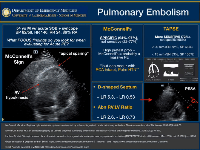
Right ventricular systolic pressure 35 mmhg is consistent the 60 60 sign has gained recent attention putatively indicating an acute cause of elevated right ventricular pressures with a pulmonary valve acceleration time 60 ms and a tricuspid regurgitation jet 30 but 60 mmhg.
Rv lv ratio pulmonary embolism. Optalyse pe optimum duration of acoustic pulse thrombolysis procedure in acute pulmonary embolism 21. Right ventricular dilatation rvd rv lv ratio 0 9 5. Right ventricular wall can be thickened 4 mm often observed in congenital heart disease or dilated in acquired heart disease free wall may be hypokinetic. A right ventricle left ventricle rv lv ratio 1 0 was not associated with fewer favorable outcomes in patients with symptomatic acute pulmonary embolism pe who were otherwise considered low risk according to study results published in the american journal of respiratory and critical care medicine.
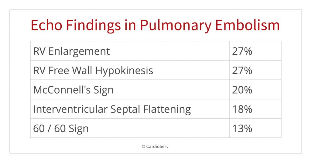
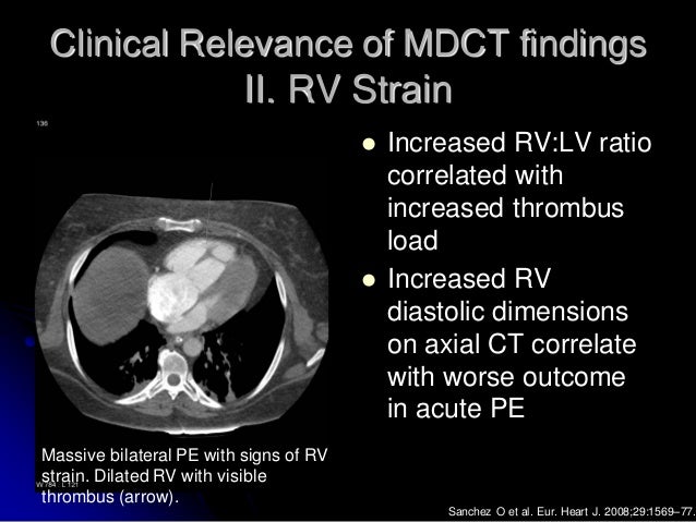
The objective of this study is to identify a clinical scenario for which normal ct derived right to left ventricular rv lv ratio. In the study by araoz et al 42 an rv lv diameter ratio greater than 1 was associated with a 3. The right ventricular to left ventricular diameter rv lv ratio measured at ct pulmonary angiogram ctpa has been shown to provide valuable information in patients with pulmonary arterial hypertension and to predict death or deterioration in acute pulmonary embolism. Rv free wall hypokinesis 27.
In this patient level post hoc analysis of 2 dutch clinical trials hestia. Epub ahead of print rv lv ratio measurement seems to have no role in low risk patients with pulmonary embolism treated at home triaged by hestia criteria. Additional studies have estimated that an rv lv diameter ratio superior to 1 5 indicates a severe episode of pe 36 39 41. A jase study in 2016 analyzed the findings from 511 consecutive patients with pulmonary embolism.
4 12 mg of tpa for 2 6 hrs. The study assessed the frequency of echo findings in pulmonary embolism with the following findings. Demographic characteristics ild subtype echocardiography and. There is variability in guideline recommendations for assessment of the right ventricle rv with imaging as prognostic information after acute pulmonary embolism pe.
Rv enlargement 27. 0 3 0 4 difference in. 24 mg of tpa. Mcconnell s sign 20.
0 42 difference in rv lv ratio. Normal ventricular diameter ratio on ct provides adequate assessment for critical right ventricular strain among patients with acute pulmonary embolism the international journal of cardiovascular imaging 32 7 2016. Elevated right ventricular pressures. Updates in echo for diagnosing pulmonary embolism.
This is best appreciated on parasternal long axis projections. May have a role in assessment. Plethoric inferior vena. Am j respir crit care med.

Eight Pearls For The Crashing Patient With Massive Pe
Pulmonary Embolism In Echo
Moderate Acute Dilatation Of The Rv In A 55 Year Old Man With Massive Download Scientific Diagram
Imaging Of Pulmonary Embolism
Pulmonary Embolism Litfl Ccc Respiratory

Right Heart Strain Radiology Reference Article Radiopaedia Org

Echocardiography In Pulmonary Embolism The Clot Thickens Pulmonary Embolism Pulmonary Pulmonary Emboli

Pin On Structure And Function

Echocardiographic Examination Of A Patient Admitted For A Recurrent Download Scientific Diagram
Peripheral Matters Current Status Of Interventional Therapies In Acute Pulmonary Embolism American College Of Cardiology

2019 Esc Guidelines For The Diagnosis And Management Of Acute Pulmonary Embolism Developed In Collaboration With The European Respiratory Society Ers European Respiratory Society

Chronic Pulmonary Embolism Diagnosis Abstract Europe Pmc

Original And Simplified Pulmonary Embolism Severity Index Download Table
Plos One Association Between Computed Tomography Obstruction Index And Mortality In Elderly Patients With Acute Pulmonary Embolism A Prospective Validation Study

Diagnostic Implications Of Computed Tomography Pulmonary Angiography In Patients With Pulmonary Embolism
Signs And Symptoms Of Pulmonary Embolism Pe Pulmonary Embolism Signs Symptoms Acutepe Diagnosis

Pdf Management Of Patients With High Risk Pulmonary Embolism A Narrative Review

Submassive Massive Pe Emcrit Project
Https Encrypted Tbn0 Gstatic Com Images Q Tbn 3aand9gcsmnit Cbktx7mceehchohiwmhirjkzwqfopwqqwwqiml8g5yti Usqp Cau

A Novel Ecg Parameter For Diagnosis Of Acute Pulmonary Embolism Rs Time Rs Time In Acute Pulmonary Embolism Sciencedirect
Saddle Pulmonary Embolus Jetem

Pert Activation Pert Indicates Pulmonary Embolism Response Team Download Scientific Diagram
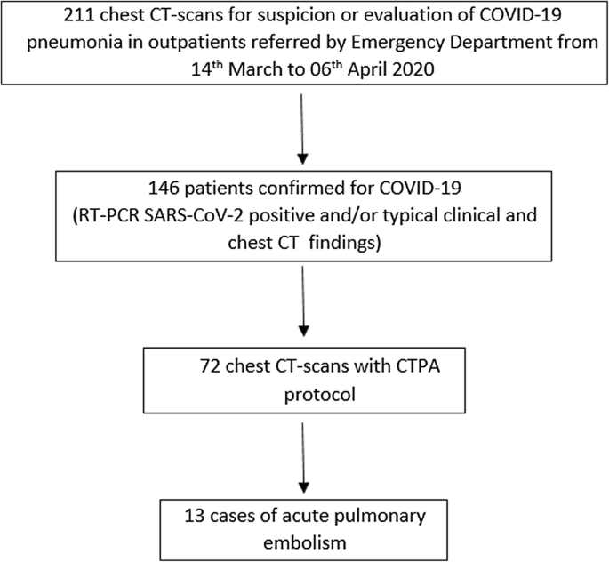
Acute Pulmonary Embolism In Non Hospitalized Covid 19 Patients Referred To Ctpa By Emergency Department Springerlink

New Horizons In Pulmonary Embolism Treatment

Ultrasound Assisted Catheter Directed Thrombolysis For Acute Intermediate Risk Pulmonary Embolism Tctmd Com

Https Encrypted Tbn0 Gstatic Com Images Q Tbn 3aand9gctdtuy2pnbihwpvwrua8qmba3h5lcihlvpnga Usqp Cau

Venous Thromboembolism Vte Management Summary Pulmonary Embolism Management Pulmonary Embolism Response

Pin By Miracleword The Rapper On Echo Pulmonary Embolism Diagnostic Medical Sonography Cardiac Sonography

Table 1 From Severe Pulmonary Embolism Pulmonary Artery Clot Load Scores And Cardiovascular Parameters As Predictors Of Mortality Semantic Scholar
Central Pulmonary Embolism In A 35 Year Old Man Ctpa Shown As Download Scientific Diagram

Https Encrypted Tbn0 Gstatic Com Images Q Tbn 3aand9gcq8z808z8jtfpc Qs6sfbgxn5dlnr9wfpcljq Usqp Cau

Assessment Of Acute Pulmonary Embolism Outcome In Hospital Through Tricuspid Annular Plane Systolic Excursion Versus Pulmonary Embolism Severity Index Score Sciencedirect

Acute Pulmonary Embolism Nejm

Gorgeous Rvot Ultrasound Humor Ultrasound Technician Medical Photos

Transverse Contrast Enhanced Ct Scan Showing Maximum Minor Axis Download Scientific Diagram

Table 2 From Submassive Pulmonary Embolism Semantic Scholar

Pdf Immediate And Late Impact Of Reperfusion Therapies In Acute Pulmonary Embolism

Winfocus 60 60 Sign Clinching The Pulmonary Embolism

Treatment Of Massive Or Submassive Acute Pulmonary Embolism With Catheter Directed Thrombolysis American Journal Of Cardiology

Apical Hypertrophic Cardiomyopathy A Lv Angiography Demonstrates Apical Hypertrophy Hypertrophic Cardiomyopathy Human Body Anatomy Diastolic Heart Failure

Frequency Of Normal Arterial Blood Gas Tests In Patients With And Download Table